Ultrasound: genital, fetal, and breast ultrasound
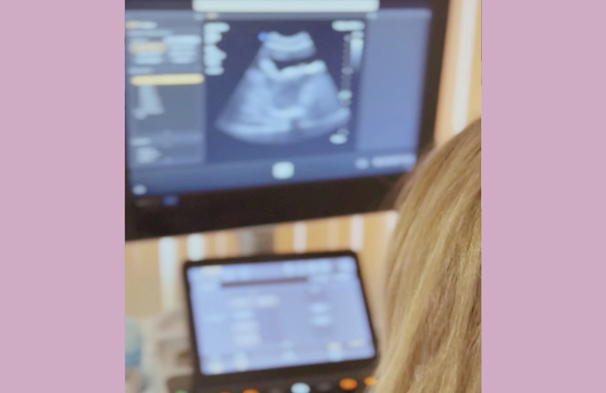
Ultrasound examinations are safe, accurate, and painless procedures that allow for real-time assessment of internal organs. The clinic performs genital (gynecological) ultrasound, fetal ultrasound (see Pregnancy Care), and breast ultrasound.
How is ultrasound performed?
The examination is conducted using ultrasound with a special sensor, which displays the image of the examined tissues on a screen. The procedure is quick, painless, and does not require complex preparation. The doctor systematically examines internal structures, assesses the shape of organs, changes, blood flow, dimensions, and possible alterations.
Genital ultrasound
Why is it performed?
Genital ultrasound is performed to assess the uterus, endometrium, ovaries, fallopian tubes, and surrounding tissues. It helps to identify cysts, fibroids, endometriosis foci, polyps, inflammatory changes, causes of infertility, or menstrual cycle disorders.
When is it necessary?
It is recommended when:
– experiencing lower abdominal pain, tension, or discomfort;
– menstrual cycle is disrupted;
– periods are heavy, painful, or irregular;
– there is unexplained bleeding or discharge;
– ovarian or uterine pathologies are suspected;
– investigating causes of infertility;
– assessing the position of an IUD;
– conducting a preventive check-up.
Breast ultrasound
Breast ultrasound helps to assess breast tissues, identify nodules, cysts, or other changes, and is often recommended as an additional examination along with mammography or as a substitute for younger women. The examination reveals the detailed structure of tissues and allows for timely detection of changes.